
Edition 7 - June, 2000
Edition 7 - June, 2000 |
THE "VANBREUSEGHEM MYCOTHEQUE" |
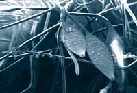 Microsporum canis IHEM 16218 (isolated from human skin) |
The BCCM/IHEM Collection has recently integrated the 12,000 fungi isolates of the "Vanbreuseghem Mycotheque", or "RV Collection", from the Institute of Tropical Medicine in Antwerp. These fungi were collected by Prof. R.Vanbreuseghem over a period of fifty years and - in effect - represent his legacy.
Vanbreuseghem was born in 1909 in Monceau sur Sambre, and graduated as doctor of medicine at the State University of Liège in 1934. After receiving his diploma from the Institute of Tropical Medicine in Antwerp, he worked from 1935 to 1946 in the former Belgian Congo. Returning to Europe, he went to Paris where he had the opportunity of working with famous mycologists such as Maurice Langeron and his friend Emile Rivalier. It was during his stay in Paris that, in 1947, he collected the first isolates of an impressive collection of 12,145 specimens from 72,000 material samples. Mainly dermatophytes at the beginning, each specimen bears an "RV" number.
The five genera of dermatophytes are well represented, with 33 Keratinomyces, 61 Epidermophyton, and 346 Trichophyton mentagrophytes isolates, but Microsporum and Trichophyton are predominant. In fact, the collection of Microsporum includes 1,289 strains distributed among 22 different species. Microsporum langeroni, an anthropophilic tropical species, frequently responsible for tinea capitis in Central Africa, comes first, followed by Microsporum canis, a worldwide zoophilic species, which is more common in developed countries.
The largest group of dermatophytes, however, comprises 1,664 Trichophyton isolates belonging to 21 different species. Among them are Trichophyton violaceum and Trichophyton soudanense, two tropical anthropophilic species often responsible for tinea capitis.These are followed by another worlwide dermatophyte, Trichophyton rubrum, with 293 isolates from all over the world.
The collection of dermatophytes also holds rarer specimens, such as a unique isolate of Microsporum vanbreuseghemii recovered from soil in New Orleans in 1961. In addition, there are some cultures of Trichophyton kuriangei - another species isolated and described by Vanbreuseghem in 1961 in the small village of Kuriange, near Bujumbura. To date, this species has never been found anywhere else.
Collections are regularly "reorganised", as - occasionally - certain isolates need to be reclassified when new species are described. With the description of Trichophyton raubitschekii for example, some of our tropical "Trichophyton rubrum" isolates had to be transferred to this new species. Some old species such as Microsporum gypseum and Microsporum langeroni are complexes and will problably be reclassified in the future. Yeasts are also regularly reclassified. Pityrosporum or Malassezia species are potentially pathogenic lipophilic yeasts living on the skin of warm-blooded animals. Traditionally, they were seen as two different species. One, Pityrosporum ovale, is related with man, and has recently been renamed Malassezia furfur, while the other Pityrosporum canis, which is more related with animals has been renamed Malassezia pachydermatis. In 1996, molecular biological techniques proved that P. ovale was a complex and had to be subdivided into six different taxa. This must be applied to the 227 P. ovale of the RV collection.
Trichosporon species are also concerned: indeed, there was one species traditionally called Trichosporum cutaneum or Trichosporum beigelii, but again, the genus was subdivided into 19 taxa, six of them associated with clinical cases. That means that the 123 Trichosporon isolates of the RV Collection must be reclassified.
The problem is different for Candida albicans isolates. This yeast is actually an endosaprophytic yeast living in the digestive tract of mammals and birds, and candidiasis of the oral mucosa is a disease recognised since antiquity. Today, this yeast has gained renewed significance as an infection often seen in AIDS patients. Until recently, the identification of this species could be performed on the basis of morphological characteristics, i.e the presence on specific media of big spores called chlamydospores. This was a cheap and quick method to identify all the C. albicans isolates of the RV Collection. But in 1995, on the basis of molecular criteria, Sullivan described a new species named Candida dubliniensis, which showed the same morphological characteristics as C. albicans.
These isolates were first considered as atypical C. albicans. The majority came from the oral cavity of AIDS-patients and seemed to develop a stable fluconazole-resistant phenotype very quickly. Moreover, it became obvious that no classical biochemical or physiological test could discriminate both species and that DNA studies should be used to reclassify the 1,055 C.albicans isolates of the RV Collection.
The RV collection of Cryptococcus neoformans is the largest, with 2,549 isolates from all over the world, and both varieties, neoformans and gattii, are well represented. Although cryptococcosis is a worldwide mycotic disease, most cases nowadays are associated with AIDS. This probably explains why so many isolates come from Central Africa where AIDS is burning: 689 isolates from Rwanda, 374 from Congo and 129 from Burundi. The first cryptococcal cases were described at the end of the nineteenth century, the diagnosis being established by direct examination of the cerebrospinal fluid which usually shows the presence of round encapsulated yeast cells. But in the sixties, F.Gatti, a physician working in Leopoldville, isolated a Cr. neoformans strain from the cerebrospinal fluid of a seven-year-old child, which showed a particular morphology in vivo: a mixture of round and elongated cells. This case was published in 1970, and the isolate described by R.Vanbreuseghem and M.Takashio was included in the RV Collection. It was forgotten until 1975, when the discovery of two different sexual reproductions corroborated the existence of two varieties Filobasidiella neoformans var. neoformans corresponding to the asexual variety neoformans, and Filobasidiella neoformans var. bacillispora corresponding to the asexual variety gattii. The type-strain is still living today and has been incorporated in the new collection. Many problems were also solved thanks to the RV collection: with the emergence of AIDS, Cr. neoformans isolates were tracked down for further studies and it could, for example, be demonstrated that, from the early fifties until 1970, the gattii variety seemed predominant in the Congo but that later, this variety disappeared from that country and gave way to neoformans which is the variety associated with AIDS.
Serotyping the African isolates, it could be demonstrated that serotype D, like the other serotypes A, B and C, could be responsible for cryptococcosis in Africa, a question that was shelved for a long time. Cr. neoformans var. neoformans and var. gattii are both exosaprophytic yeasts from the environment. The neoformans variety however is most often recovered from soil mixed with pigeon droppings, which constitute the main source of contamination for patients. Apart from isolates from clinical cases, the RV collection also includes 640 strains from diverse saprophytic origins and indeed also from pigeon droppings. Moreover, some isolates were found in Africa from dust collected in houses of AIDS and non-AIDS-associated cryptococcosis patients. This is thus the ideal material to compare strains from patients with the isolates recovered from their environment.
The collection also includes many other fungi responsible for mycoses, such as several isolates of Histoplasma duboisi, the agent of African histoplasmosis, a species described by Vanbreuseghem and named after Prof. A. Dubois, director of the Institute of Tropical Medicine in the fifties. There are also, among many others, more than a hundred Sporothrix schenkii isolates, the sporotrichosis agent, sent by South American colleagues.
There are, unfortunately, no isolates of Rhinosporidium seeberi, the rhinosporidiosis agent, nor Loboa loboi, responsible for lobomycosis, simply because both organisms cannot be maintained in culture and are only known as parasites.
In conclusion, this collection is not only a witness of the past, but is biological material for the present and the future. It is part of world heritage.
ContactHome |
Contents Edition 7 - June, 2000 |
Next Article Edition 7 - June, 2000 |